The primary objective of this project is to explore the application of deep learning in the healthcare sector, specifically through the use of transfer learning with pretrained models such as ResNet18 for the classification of X-ray images. The goal is to develop a model capable of accurately distinguishing between normal and pneumonia-affected images.
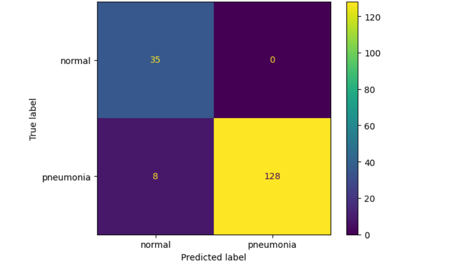
Utilizing a ResNet18 pretrained model and a dataset from Kaggle, the model exhibited strong training performance but achieved a modest validation accuracy of 65%, likely due to the limited dataset of 20 data points used for validation. Despite this, the model demonstrated excellent performance on the test set with an accuracy of 95%.
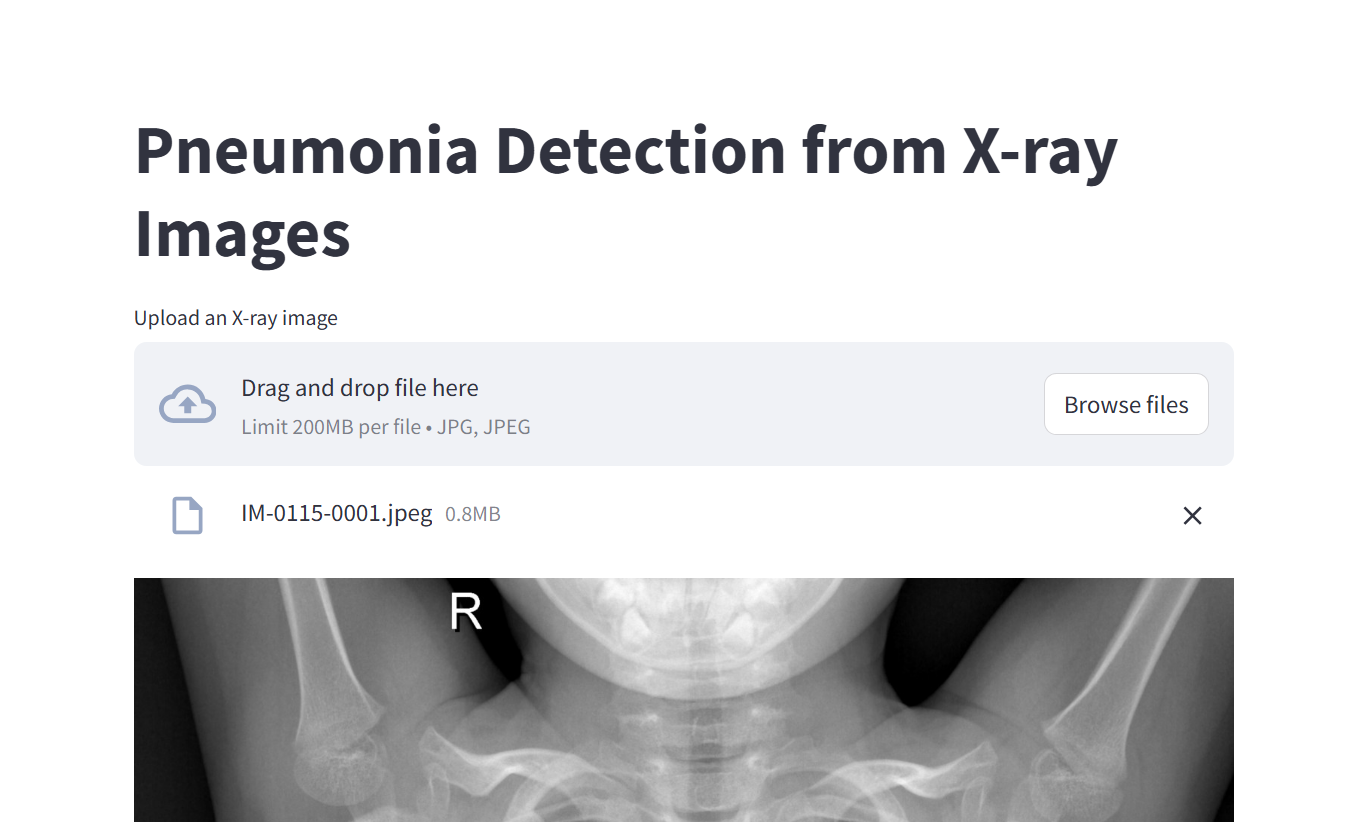
Additionally, the project includes the development and deployment of a user-friendly application on Stream lit, enabling users to upload X-ray images and receive predictions, thereby showcasing the practical utility of the model.
The “Deep Learning for Pneumonia Detection Using X-ray Imaging” project aims to demonstrate the critical role of Artificial Intelligence (AI) in healthcare, specifically in detecting diseases using historical data to train models. These models can significantly aid medical professionals in saving lives and developing strategies to reduce infections such as pneumonia. While the classification approach showcased in this project is focused on healthcare, it is versatile and can be applied in various other sectors including finance, sports, and education.
To overcome resource limitations, such as the lack of GPU-enabled computers, transfer learning with pretrained models like ResNet18 was utilized. The training was conducted on Google Colab, which offers free but limited GPU services. Leveraging pretrained models, trained under optimal conditions, ensures high accuracy—a crucial requirement for health-related outcomes.
This project aims to leverage deep learning techniques for the classification of pneumonia from chest X-ray images. By utilizing advanced convolutional neural networks (CNNs), the study seeks to enhance diagnostic accuracy and support medical professionals in detecting pneumonia efficiently.
The methodology involves data preprocessing, model training using CNN architectures, and performance evaluation on a labeled dataset of X-ray images. Key steps include data augmentation, hyperparameter tuning, and cross-validation to ensure robustness. Below are some of the processes:
import os

import numpy as np
import pandas as pd
from PIL import Image

from tqdm.auto import tqdm
from matplotlib import pyplot as plt
from sklearn.model_selection import train_test_split
from sklearn.metrics import ConfusionMatrixDisplay, confusion_matrix


import torch
from torchinfo import summary
import torch.nn.functional as F
from torch.optim.lr_scheduler import StepLR
import torch.nn as nn
from torch.optim import Adam
from torch.utils.data import Dataset, DataLoader, random_split
from torchvision import transforms, datasets
from torchvision import models
from torchvision.models import resnet18
# define data directories
train_path = ("/content/drive/MyDrive/chest_xray/train")
test_path = ("/content/drive/MyDrive/chest_xray/test")
val_path = ("/content/drive/MyDrive/chest_xray/val")
# get classes from the training data
classes = os.listdir(train_path)
print('There are', len(classes), 'classes')
print(classes)
# converting images to RGB
class ConvertToRGB():
  def __call__(self, img):
    if img.mode != 'RGB':
      img = img.convert("RGB")
    return img

# defining transforms

transform = transforms.Compose(
    [
        ConvertToRGB(),
        transforms.Resize((224, 224)),
        transforms.ToTensor(),


    ]
)
# creating  data sets using ImageFolder
train_dataset = datasets.ImageFolder(root = train_path, transform = transform)
val_dataset = datasets.ImageFolder(root = val_path, transform = transform)
test_dataset = datasets.ImageFolder(root = test_path, transform = transform) # to be used for testing

print(train_dataset)
print(val_dataset)
# introducing a pretrained model
model = models.resnet18(weights= models.ResNet18_Weights.DEFAULT)
print(model)
# training function

def train_model(model, train_loader, val_loader, loss_fn, optimizer, num_epochs, scheduler, early_stopping, checkpointing,   device = 'cpu' ):
    for epoch in range(num_epochs):
        print(f"Epoch {epoch+1}/{num_epochs}")
        print('-' * 10)

        # Iterate over training and validation phases
        for phase in ['train', 'val']:
            if phase == 'train':
                model.train()  # Set model to training mode
                data_loader = train_loader
            else:
                model.eval()  # Set model to evaluation mode
                data_loader = val_loader

            running_loss = 0.0
            running_corrects = 0

            # Iterate over data
            for inputs, labels in data_loader:
                inputs, labels = inputs.to(device), labels.to(device)

                optimizer.zero_grad()

                # Forward pass
                with torch.set_grad_enabled(phase == 'train'):
                    outputs = model(inputs)
                    _, preds = torch.max(outputs, 1)
                    loss = loss_fn(outputs, labels)

                    # Backward pass + optimization only in training phase
                    if phase == 'train':
                        loss.backward()
                        optimizer.step()

                # Statistics
                running_loss += loss.item() * inputs.size(0)
                running_corrects += torch.sum(preds == labels.data)

            epoch_loss = running_loss / len(data_loader.dataset)
            epoch_acc = running_corrects.double() / len(data_loader.dataset)

            print(f'{phase}_Loss: {epoch_loss:.4f} {phase}_Acc: {epoch_acc:.4f}')
#load the model and evaluate the test data
model.load_state_dict(torch.load("pneumonia_model.pth", weights_only = True))

model.to(device)

model.eval()

y_true, y_pred  =[], []
with torch.no_grad():
    for images, labels in test_loader:
        images, labels = images.to(device), labels.to(device)
        outputs = model(images)
        _, preds = torch.max(outputs,1)

        y_true.extend(labels.cpu().numpy())
        y_pred.extend(preds.cpu().numpy())

        accuracy  = accuracy_score(y_true, y_pred)

print(f'The model accuracy on unseen data  is', accuracy)
The model accuracy on unseen data is 0.9532163742690059
disp = ConfusionMatrixDisplay(matrix_test, display_labels = ['normal', 'pneumonia'])
disp.plot()

def test_prediction(model, dataset,i):
    image = dataset[i][0]
    image_batch= image.unsqueeze(0)

    model = model.to(device)

    with torch.no_grad():
        image_batch = image_batch.to(device)
        output = model(image_batch)
        _, predicted_class = output.max(1)

        # mapping the predcition to the class
        class_names = ['normal', 'pneumonia']

        # Map predictions to class names
        predicted_labels = [class_names[idx] for idx in predicted_class.cpu().numpy()]  # Convert to list of
        class names

    #Print results
    for idx, label in enumerate(predicted_labels):
          print(f"The predicted label for image {i + 1} is '{label}'.")

          # displaying the image
          image = np.transpose(image, (1, 2, 0))
          image = np.clip(image, 0, 1)
          plt.imshow(image)
          plt.axis('off')
          predicted_label = predicted_labels[0]
          plt.text(10, 20 , f'Predicted: {predicted_label}', fontsize = 15, color = "red", bbox=dict(facecolor=
           'white',
          alpha=0.7))
          plt.show()

          predicted_labels =class_names[predicted_class.item()]

          print('The predicted label is ', predicted_labels)
Pnuemonia-Xray-ImageClassification
The findings indicate that deep learning models, particularly CNNs, can effectively analyze X-ray images to detect pneumonia. Challenges such as dataset bias and model interpretability are discussed, along with potential improvements for future research.
This study demonstrates the potential of deep learning in medical imaging, particularly for pneumonia detection. By integrating AI tools into healthcare, diagnostic efficiency and accuracy can be significantly improved, paving the way for advanced medical solutions.
This report documents the development and deployment of a Streamlit application for pneumonia detection using deep learning models. The project utilizes ResNet-18, a convolutional neural network (CNN), to classify X-ray images as either "Pneumonia" or "Normal." The application aims to provide a user-friendly interface for medical professionals to quickly and accurately assess chest X-rays.
The application integrates a pre-trained ResNet-18 model, fine-tuned for binary classification. Key stages include:
ResNet-18 is loaded with pretrained weights (ResNet18_Weights.DEFAULT) for feature extraction.
The fully connected layer is customized for binary classification with softmax activation.
Input images are converted to RGB (if necessary).
Images are resized to 224x224 pixels and normalized using standard ImageNet mean and standard deviation values.
Uploaded X-ray images are processed through the neural network to output probabilities.
The model determines the class label (Normal or Pneumonia) based on the highest probability.
Visualization:
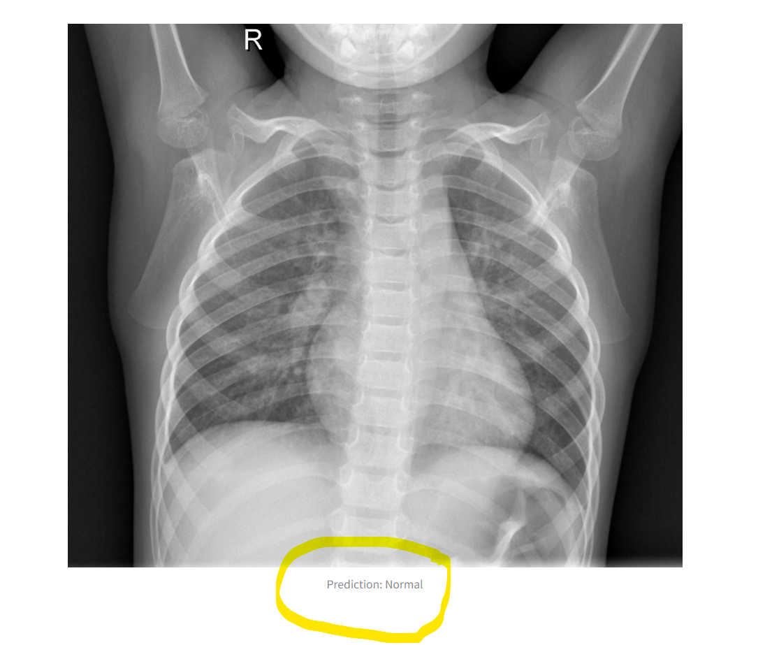
The original image is displayed alongside the prediction result.
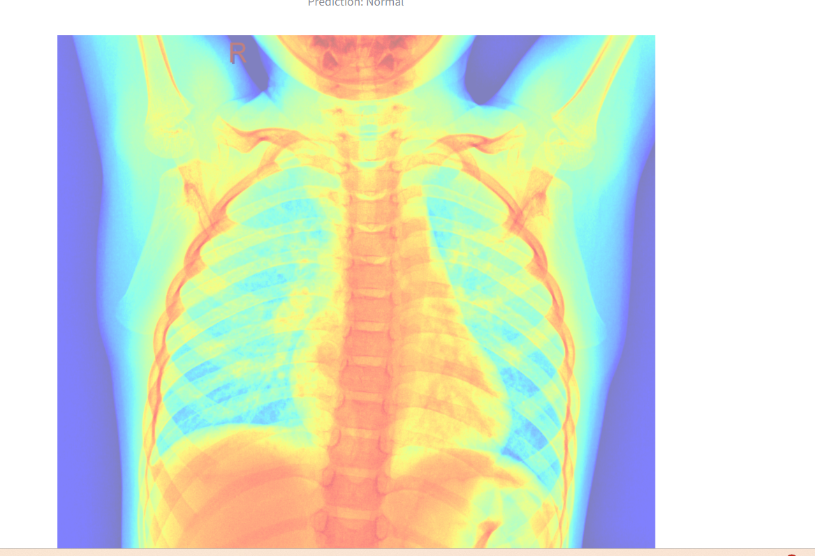
A heatmap overlay is generated for better interpretability.
The application demonstrates reliable performance on test images. It accurately distinguishes between "Normal" and "Pneumonia" cases when provided with valid X-ray images. Results are displayed directly in the Streamlit interface, making it accessible for non-technical users. See the image below:

The app interface

The output image and label

The heatmap overlay
Ease of Use: The Streamlit interface simplifies interaction, allowing users to upload images and receive instant predictions.
Performance: Leveraging a fine-tuned ResNet-18 model ensures robust feature extraction and classification accuracy.
Visualization: The app enhances interpretability with a heatmap overlay.
Generalization: The model’s performance is reliant on the quality and diversity of the training dataset.
Device Limitation: Computations were performed on a CPU, which might affect processing time for large datasets.
Expanding the dataset to improve generalization across diverse X-ray imaging conditions.
Incorporating explainability tools such as Grad-CAM for enhanced interpretability.
This Streamlit application effectively applies deep learning to classify pneumonia from X-ray images. By offering a straightforward interface and reliable predictions, it demonstrates the practical potential of AI in healthcare diagnostics.